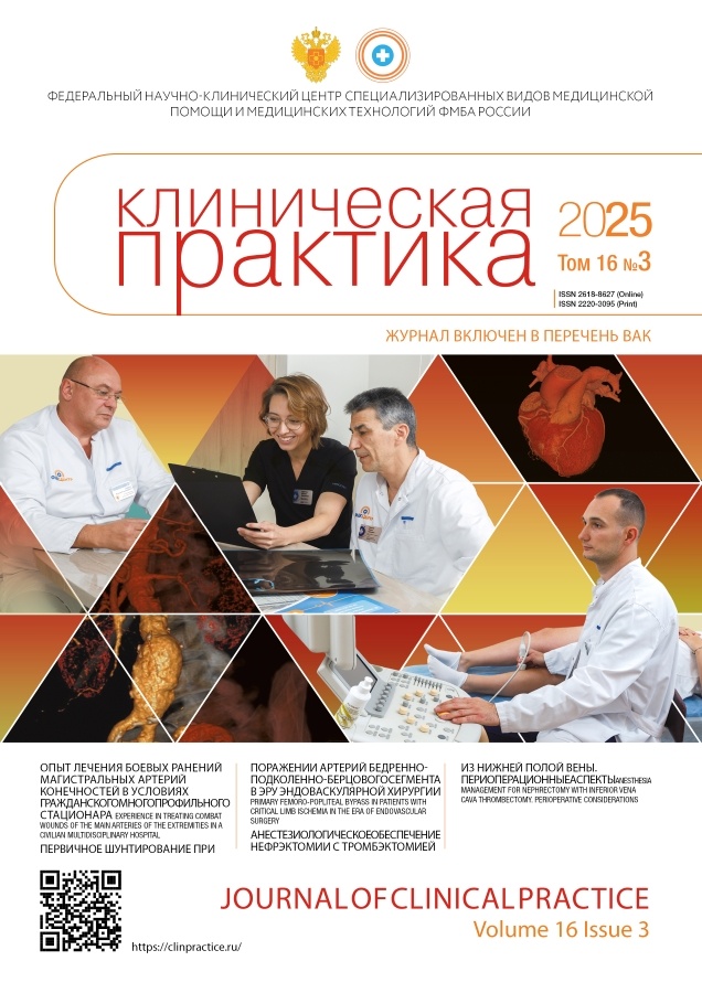
Journal of Clinical Practice
Quarterly peer-review medical journal.
Editor-in-chief
- professor Alexandex I. Troitsky, MD, Dr. Sci. (Medicine)
ORCID: 0000-0003-2143-8696; Scopus Author ID: 57202735847
Main handling editor
- Vladimir P. Baklaushev, MD, Dr. Sci. (Medicine)
ORCID: 0000-0003-1039-4245; Researcher ID: H-2426-2013
Publisher
- Eco-Vector Publishing group
Founder
- Federal Research Clinical Center FMBA of Russia
WEB: https://fnkc-fmba.ru
About
The main idea of our journal is to provide description and analysis of clinical cases with severe, rare and difficult for diagnoses diseases, occurred in the clinics of Federal Medical-Biological Agency of Russia. Such clinical analysis is aimed to develop “clinical” type of thinking, always have been the characteristic feature of Russian/USSR medical school. The journal purpose is also to improve scientific discussions and cooperation between physicians of different specialties.
Revival of historical traditions in our journal is the one of the components of continuing education, which is especially important in “closed” territories, where doctors can`t regularly participate in clinical conferences. An important aspect is to provide a printed tribune for any doctor who has an interesting clinical observation and wish to share his experience with colleagues. That is why we named our journal "Clinical Practice" and address it, first of all, those skilled in applied medicine. Of course, we also publish the results of original researches, clinical guidelines, current reviews and medical news. The journal is multidisciplinary and we hope that it will be interesting to doctors of different specialties. The journal is published by means of the Federal Research and Clinical Center of FMBA of Russia. Placement of all materials, except for advertising, are free of charge to authors.
Types of accepted articles
- reviews;
- systematic reviews and meta-analysis;
- original study articles;
- case reports and series of cases;
- letters to the editor;
- hystorical articles
The joutnal accept manuscripts in English and in Russian.
Publication, distribution and indexation
- Russian and English full-text articles;
- issues publish quarterly, 4 times per year;
- no obligatory APC, Platinum Open Access
- articles distribute under the Creative Commons Attribution-NonCommercial-NoDerivates 4.0 International License (CC BY-NC-ND 4.0).
Indexation
- Russian Science Citation Index (elibrary.ru)
- DOAJ
- CrossRef
- Dimensions
- Google Scholar
- Ulrich's Periodicals Directory
- CyberLeninka
Current Issue
Vol 16, No 3 (2025)
- Year: 2025
- Published: 26.10.2025
- Articles: 12
- URL: https://clinpractice.ru/clinpractice/issue/view/10215
The experience of treating battle injuries of the magistral arteries in the limbs in the settings of the civilian multi-profile in-patient hospital
Abstract
BACKGROUND: The modern military conflict is characterized by a significant number of wounded with the damage of the magistral arteries in the limbs. Such an injury is accompanied by the possibility of lethal outcome and by the high risk of limb amputation. The treatment of the injuries in the major arteries requires high qualification of the medical staff and sufficient equipment basis. The optimal tactics for this still remains the matter of discussion. AIM: to define the specific features of the surgical tactics in cases of injured magistral arteries in the settings of the civilian specialized in-patient hospital in the regions adjacent to the scene of military operations. METHODS: The analyzed data included the treatment results in 57 patients with battle injuries of the magistral arteries in the limbs, in which we have managed to track the direct result of restoring the arteries within not less than two days. The variety of manifestations observed in cases of injuries was demonstrated using 8 clinical cases. The surgical tactics was defined by the degree of ischemia in the muscles and the extent of damaging the tissues in the limb. Amputations were conducted in cases of developing the ischemic contracture or in cases of significantly damaged limb tissues. RESULTS: The resection of the artery with autovenous prosthetic replacement was done in 49 cases, while the circular resection of the artery with the direct anastomosis — in 8 cases. Within the earliest post-surgery period (first two days) due to the post-ischemic syndrome, the usage of the extracorporeal detoxication methods was required in 5 (9%) wounded. The restoration of the peripheral circulation was observed in 56 (98.2%) cases, the secondary amputation of the lower limb was done only in 1 (1.8%) operated patient. No fatal outcomes were reported (0%). CONCLUSION: In the modern military conflict, the battle contact line can be located in the direct proximity from the well-equipped civilian healthcare institutions, at the premises of which the high-tech medical aid is accessible. Our experience shows that, in case of performing the complex surgeries, the follow-up within the early period is practicable to be organized at the site with avoiding the immediate evacuation. In cases of damaging the magistral artery in the limb, the main parameter affecting the possibility of saving the limb itself, is the degree of ischemia in the muscles. The irreversible ischemia is often hard to define and the development of the ischemic contracture should be taken as the guidance. The time of injury, the absence of pulse, of the active movements or sensitivity cannot serve as an indication for amputation. The algorithm developed by us has shown its high efficiency.
 7-22
7-22


Original Study Articles
Risk factors for post-operative cognitive dysfunction in neurosurgical patients
Abstract
BACKGROUND: The impact of various risk factors on the development of post-operative cognitive dysfunction in neurosurgical patients requires research for decreasing the probability of developing this complication. AIM: To determine the effects of extra- and intraoperative risk factors on the development of post-operative cognitive dysfunction in neurosurgical patients after undergoing a vertebral column surgery with long-running anesthetic support. METHODS: The research was carried out among the neurosurgical patients with previous surgical intervention in the vertebral column, within the premises of the Neurosurgery Department of the State Budgetary Healthcare Institution of the Tyumen Oblast “Regional Clinical Hospital No. 2”. The evaluation included the cognitive functions before surgery and on Day 3 after the surgical intervention using the Montreal Cognitive Assessment (MoCA), along with a panel of Isaac tests and the Munsterberg test. The calculated coefficients were the Pearson’s and the point biserial correlation coefficients regarding the following intraoperative risk factors: type and duration of anesthetic management, medications used for anesthesia and muscle relaxation, as well as the type of surgery. Evaluations were also made for the interrelation between the development of post-operative cognitive dysfunction and the following extra-operational risk factors: the age, the body mass index, the number of education years, the presence of arterial hypertension or diabetes and smoking. RESULTS: A notable positive correlation was observed between the development of post-operative cognitive dysfunction and the age (r=0.53; p <0.01), moderate correlation with the body mass index (r=0.35; p <0.01) and with the presence of arterial hypertension (r=0.42; p <0.05). A moderate negative relation was observed for the number of education years and the development of post-operative cognitive dysfunction (r=-0.36; p <0.01). The relation of the presence of diabetes with post-operative cognitive dysfunction did not show significant correlation. Smoking and surgery duration show low level of interrelation, which does not allow to comprehensively interpret the obtained results as significant. The type of surgical intervention and the duration of anesthetic support did not correlate with the development of post-operative cognitive dysfunction (r <0.1; p <0.01). A moderate correlation was found for the anesthesia conducting using a drug combination of desflurane+fentanyl (r=0.31; p <0.05) along with the mild one when combining sevoflurane+fentanyl+ketamine (r=0.25; p <0.05). The usage of fentanyl together with sevoflurane (r=0.07), propofol (r=-0.1) and sodium oxybutyrate (r=0.05) does not lead to post-operative cognitive dysfunction (p <0.05). CONCLUSION: Elderly age, high body mass index, presence of arterial hypertension and low education level increase the risks of developing post-operative cognitive dysfunction. Using the desflurane+fentanyl and sevoflurane+fentanyl+ketamine combinations can also contribute to the occurrence of cognitive disorders.
 23-29
23-29


Primary femoro-popliteal-tibiofibular bypass in patients with critical limb ischemia in the era of endovascular surgery
Abstract
BACKGROUND: In the majority of patients with critical ischemia in the lower limbs, the findings include the «multi-level» atherosclerotic lesions in the arteries of the femoral-popliteal-tibiofibular segment. The optimal method of re-vascularisation in this cohort of patients is not defined as of today. AIM: To evaluate the efficiency of conducting the initial autovenous tibiofibular bypass surgery in case of lesions in the arteries of the femoral-popliteal-tibiofibular segment in patients with critical ischemia of the lower limbs. METHODS: The analysis included the results of the initial tibiofibular autovenous bypass surgeries, performed in 112 patients at the Federal State Budgetary Institution «Federal Clinical Center of High Medical Technologies» under the Russian Federal Medical-Biological Agency during the period from 2010 until 2021, of which 25 (22.3%) individuals had the stage III chronic arterial insufficiency in the lower limbs, 87 (77.7%) — stage IV acc. to the Fountain–Pokrovsky classification. The distribution by the atherosclerotic lesion in arteries of the lower limbs with taking into consideration the TASC II classification was the following: type C — in 9 (8.0%), type D — in 103 (92.0%). RESULTS: Within the 30 days period, 4 (3.6%) patients have shown the presence of unfavorable cardio-vascular events, 3 (2.7%) cases resulted in the early high amputation. The perioperative mortality rate was 2.7% (n=3). The primary passability of the tibiofibular autovenous bypass was 91%, 76% and 67% in 1, 3 and 5 years, while the secondary passability was 93%, 80% and 71%; the limb survival rate was 98%, 86% and 81,5%; the overall survival of the patients was 88.5%, 81% and 70%, respectively. CONCLUSION: The initial tibiofibular autovenous bypass surgeries (bypass first) represent the effective and safe method of surgical treatment for atherosclerotic lesions in the arteries of the femoral-popliteal-tibiofibular segment in patients with critical ischemia of the lower limbs. Open-access surgeries in the era of endovascular surgery can be used as the first line therapy with comparable direct and remote results.
 30-37
30-37


The tactics of weaning from cardiopulmonary bypass with blood-saving technique in cardiac surgery
Abstract
BACKGROUND: Cardiac surgery under cardiopulmonary bypass is typically characterized by significant blood loss and the need for donor red blood cell transfusions. In addition to the inflammatory response, hemodilution, hypocoagulation, and blood loss significantly contributes to the development of perioperative anemia associated with the weaning from the cardiopulmonary bypass. AIM: Optimization of weaning from cardiopulmonary bypass to reduce blood loss during cardiac surgery. METHODS: Patients undergoing cardiac surgery under cardiopulmonary bypass (n=62) were divided into two groups. In the study group (n=31), all blood from cardiopulmonary bypass circuit was returned to the patient's central vein at the end of the cardiopulmonary bypass. In the comparison group (n=31), a standard method of pushing a residual blood volume from the cardiopulmonary bypass circuit with normal saline was used. Laboratory and instrumental data were analyzed. RESULTS: Intraoperative blood loss in the study group was significantly lower than in the comparison group (500 [470–520] ml versus 800 [760–830] ml, p=0.0001). Twenty-four hours after surgery, creatinine, alanine aminotransferase, and amylase concentrations were higher in the study group than in the comparison group. At the end of surgery, the study group also had higher cardiac index (3.1 [2.8–3.6] versus 2.8 [2.6–3.1] l/m2 per minute, p=0.018) and global ejection fraction (28 [22–31] versus 22 [19–24]%], p=0.011). No adverse events or reactions were registered during the study. CONCLUSION: Complete blood return after cardiopulmonary bypass results in higher hemoglobin and hematocrit levels in the early postoperative period, accompanied by less blood loss and higher cardiac index and global ejection fraction after the main stage of the surgery without significant adverse events.
 38-46
38-46


Reviews
The anesthetic management and the specific features of perioperative management in cases of nephrectomy with thrombectomy from the inferior vena cava in patients with renal cell cancer
Abstract
Renal cell cancer is one of the most widespread oncourological diseases (90% of all the malignant neoplasms in the kidneys) with high mortality. Every year worldwide, approximately 120,000 new cases of renal cell cancer are diagnosed, which is approximately 2% within the structure of the cancer incidence rates, and 65% of the patients are being diagnosed in the developed countries. Nephrectomy is the main method of radical therapy for such patients. In cases of tumor thrombosis of the inferior vena cava, which develops in 25–30% of the cases of renal cell cancer and represents a lethal complication of this disease due to the fragmentation of the thrombotic masses and developing pulmonary embolism, nephrectomy with thrombectomy is indicated. A special category includes the patients with renal cell cancer, complicated by the tumor thrombosis of the inferior vena cava with grades III (thrombus located at the level or above the hepatic veins, but below the diaphragm) and IV (thrombus spreading into the supradiaphragmatic inferior vena cava or into the right atrium) according to the classification by the Mayo Clinic, in which the surgical strategy is accompanied by significantly traumatic manipulations with the liver, the suprahepatic segment of the inferior vena cava, as well as with the heart chambers, suggesting the parallel cardiosurgical intervention. Surgical interventions with this background are accompanied by the complete or the parallel methods of extracorporeal circulation. The initially burdened status of the patient (tumor-related intoxication, anemia, hyperazotemia, in a number of cases thrombosis of the venous system in the lower limbs along with the concomitant abnormalities) and the extent of surgical intervention determine the high risk of complications (up to 93%) and hospital mortality (up to 10%). The preoperative evaluation of the risks of surgery, defining the most favorable tactics for the patient and the thorough preoperative preparation are necessary for the safest course of surgery and for the early rehabilitation of the patient. Currently, there is no unified commonly accepted algorithm adopted for managing such patients, while the developed commonly available standards often have a generalized type, not reflecting the specific features found in the patients with tumor thrombosis of the inferior vena cava. This review attempts to compile the specific features of the anesthetic management in cases of nephrectomy with thrombectomy in patients with renal cell cancer, to describe the main pathophysiological features of the tumor thrombosis of the inferior vena cava, the complications of the perioperative period, the methods for their prevention and treatment. The main directions were provided for the combined diagnostics and treatment, special attention was paid to the multi-disciplinary (urologists, oncologists, cardiovascular and cardiosurgery specialists, anesthesiologists and intensivists) team-based approach to perioperative management of the patients with tumor thrombosis of the inferior vena cava.
 47-57
47-57


Microcystic macular edema: clinical significance and pathogenetic mechanisms
Abstract
Microcystic macular edema represents a specific type of intraretinal cystic changes, localizing predominantly in the inner nuclear layer and detectable using the optical coherence tomography. Contrary to the classic concepts on the macular edema as a result of vascular permeability, microcystic macular edema is not accompanied by exudation and it is perceived as the manifestation of neuroglial dysfunction, often associated with the damaging of the optic nerve. Initially described in patients with multiple sclerosis, microcystic macular edema was subsequently detected in the wide spectrum of diseases, including glaucoma, neuromyelitis optica spectrum disorders, diabetic retinopathy, occlusion of the retinal veins, senile macular degeneration and epiretinal membranes. The key pathogenetic mechanisms are considered the retrograde transsynaptic degeneration of the ganglionic cells in the retina and the functional/structural damage of the Muller’s cells, in particular, the impaired operation of the AQP4 aquaporin channels. The morphological features of the microcystic macular edema, its location and clinical significance vary depending on the main disease and in a number of cases can act as the early biomarker of the neurodegenerative process. The article contains the pathophysiological models, the clinical correlates and the modern methods of the diagnostics of microcystic macular edema with special emphasis on the role of multimodal visualization and artificial intelligence technologies. Taking into consideration the rates of accidental detection and the potential relation to the systemic diseases, microcystic macular edema should be considered not as an isolated ophthalmology condition, but as the component of wider neuroretinal disorder requiring interdisciplinary approach to the diagnostics and follow-up.
 58-70
58-70


Differential diagnostics of non-small-cell and small-cell lung cancer: modern approaches and promising technologies
Abstract
Lung cancer represents a heterogeneous group of malignant neoplasms, among which two main forms can be distinguished — the non-small-cell and the small-cell lung cancer. These subtypes significantly differ by the histological, the molecular-genetic and the clinical characteristics, which defines the necessity of precise differential diagnostics for selecting the optimal treatment tactics. The review highlights the modern methods of diagnostics for the non-small-cell and the small-cell lung cancer, including the instrumental diagnostics, the histological and immunohistochemical examinations. Special attention was paid to the pros and cons of the promising non- and minimally invasive approaches, such as the analysis of circulating tumor cells, of the extracellular DNA, of the miRNA, of the marker proteins, of the volatile organic compounds and of the modern medical visualization (radiomics). Despite the significant progress in developing new diagnostic approaches, the problems remain that are related to the heterogeneity of tumors, the limited accessibility of the materials of small-cell lung cancer and the necessity of standardizing the new methods. The promising direction seems to the integration of multimodal approaches, combining the fluid biopsy, radiomics and the algorithms of machine learning, which can increase the precision of diagnostics and optimize the personalized treatment of the patients with various subtypes of lung cancer.
 71-87
71-87


Comorbidity background and rehabilitation potential among the cerebral stroke patients
Abstract
Stroke is one of the most significant social problems due to the high incapacitation rates among the patients. The rehabilitation of the patients in the older age group with stroke consequences is complicated by the fact that they almost always have a comorbidity background, influencing the efficiency of restoring the lost functions and the possibilities of using any technologies of medical rehabilitation. Comorbidity makes its contribution to the development of repeated stroke and plays a significant role when drafting the rehabilitation program. The review analyzes the data from scientific literature on the effects of concomitant diseases on the rehabilitation potential of patients after a past acute cerebrovascular accident. An analysis was carried out for the literature data using three data bases (PubMed, MEDLINE and eLIBRARY) for the period from 2000 until 2025 with 435 scientific articles analyzed, and for the detailed analysis, 35 publications were selected that meet the inclusion criteria. Based on the analysis conducted, various options were presented for evaluating the rehabilitation potential and for interpreting the evaluation results with taking into consideration the effect of various diseases, most frequently seen in patients with acute cerebrovascular accident. A discussion is presented on the necessity of compiling a single unified method of determining the rehabilitation potential. The analysis of literature data has shown that evaluating the comorbidity is one of the important components of the rehabilitation potential in the patient after the stroke. Determining the most significant factors shaping the rehabilitation potential in such patients is a top priority task determining the choice of rehabilitation therapy tactics and its efficiency.
 88-97
88-97


Case reports
FXTAS (Fragile X-Associated Tremor/Ataxia Syndrome)
Abstract
BACKGROUND: FXTAS Syndrome (Fragile X-Associated Tremor/Ataxia Syndrome) is a neurodegenerative disease with late onset, which manifests in men and women carrying the mutation in the FMR1 gene, located in the X-chromosome. The disease manifests with high phenotypic variability (tremor, cerebellar ataxia, parkinsonism, oculomotor disorders, cognitive and mental disorders). Due to the insufficient awareness among the physicians on this disease, FXTAS Syndrome patients often get incorrect diagnosis (essential tremor, Parkinson disease, multisystem atrophy, spinocerebellar ataxia etc.). CLINICAL CASE DESCRIPTION: The case presented is the patient aged 68 years with a severe past medical history (ischemic heart disease, post-infarction cardiosclerosis with the formation of post-infarction aneurism, arterial hypertension, pulmonary tuberculosis, chronic obstructive pulmonary disease, type 2 diabetes), in which the FXTAS Syndrome has first manifested with tremors, impaired coordination of motions, balance problems, cognitive disorders and affective disorders. The disease was confirmed by the genetic test (in the FMR1 gene, 96 CGG repeats were found). The patient’s daughter was examined with detecting the premutation of the FMR1 gene, while the grandson has a Martin-Bell syndrome. CONCLUSION: Neurologists and specialists of adjacent fields should keep in mind the FXTAS Syndrome (Fragile X-Associated Tremor/Ataxia Syndrome) and, in case of the patient having the corresponding symptoms, should rule out this rare neurodegenerative disease by arranging the genetic testing to reveal the mutation in the FMR1 gene.
 98-104


Bilateral tibial lesions as an onset of the diffuse В-cellular non-Hodgkin lymphoma
Abstract
BACKGROUND: Diffuse В-cellular non-Hodgkin lymphoma is the most widespread in this group of diseases, causing the highest number of lymphoma-related lethal outcomes worldwide. The clinical-pathological heterogeneity of the nosology negatively affects the possibilities of precise and timely diagnostics. The variety of extranodal locations of the disease requires the participation of a multidisciplinary team of high qualification specialists, the use of high-tech methods for diagnostics and the aggressive therapy. The article presents a discussion on the occurrence of the diffuse В-cellular non-Hodgkin lymphoma, on the rates of an onset with the lesions in the skeleton, on the specific features of the disease course that is mimicking the rheumatic disease, as well as on the difficulties of recognizing it due to the absence of specific symptoms at the beginning of the disease and to the necessity of using high-tech instrumental diagnostic methods. Clinical case description: The article presents a clinical case of the patient aged 57 years, hospitalized to the Regional Tuberculosis Dispensary with the provisional diagnosis of tuberculosis-associated arthritis of the ankle joints. At the out-patient phase, the differential diagnostics was conducted between the inflammatory disease of the joints, the trauma, the bone tuberculosis and the orthopedic abnormalities. The radiology image of the chest cavity organs showed the presence of diffuse pneumosclerosis, according to the data from multispiral computed tomography, a cystic transformation was found along with the space-occupying mass lesions and osteolysis in the distal areas of both tibial bones. Based on the results of the histological examination of the lower third of the left tibia, a diagnosis of diffuse В-cellular non-Hodgkin lymphoma with high proliferative activity was set, confirmed at the specialized medical institution using the method for calculating the IPI prognostic index (International Prognostic Index). Conclusion: The presented case demonstrates the necessity for oncological alertness and for multidisciplinary approach for ruling out the primary and the secondary non-Hodgkin lymphomas of bone tissue location, mimicking the bilateral disease of the joints, for the purpose of early diagnostics and timely treatment, improving the prognosis for patients with this group of diseases.
 105-111


The first experience of conducting the anesthetic support during a simultaneous surgery in a Kindler syndrome patient
Abstract
BACKGROUND: Kindler syndrome is a rare autosomal recessive disease, one of the forms of congenital epidermolysis bullosa. Clinically, the disease manifests by the development of bubbles on the skin and mucosal membranes with further cicatrization, as well as by the development of narrowing in the esophagus, the urethra, the vagina and the urinary ducts. In the presented clinical case of the first ever in Russia conducted simultaneous surgery in the settings of general combined anesthesia in a patient with Kindler syndrome, evaluation was carried out for the criteria of difficult airways, special attention was paid to the method of conducting the endotracheal intubation, to monitoring the vital functions of the organism and to following the multimodal analgesia principle. Due to the high risk of post-operative nausea and vomiting, a necessity was justified for increasing the antiemetic effect. CLINICAL CASE DESCRIPTION: The main indications to conducting the surgery in a female patient aged 49 years with congenital epidermolysis bullosa (Kindler syndrome) were the complaints of significant difficulties and pain upon swallowing, decreased appetite, presence of dysphagia with a background of inhomogeneous circular narrowing of the esophagus, pain when moving the eyes and the absence of nasal breathing with a background of nasal vestibule atresia. The main tasks of surgical treatment were the elimination of incapacitating complications and improving the quality of life for the patient. The multiplicity of stages in the treatment process was deemed impractical due to the necessity of conducting three anesthetic support procedures with high risk of additional damaging the oropharynx and upper airways during tracheal intubation, due to which, a decision was drawn up on arranging a simultaneous surgical treatment of complications of the main disease. The duration of surgery was 195 minutes, while the anesthesia lasted for 210 minutes. The performed procedures included the elimination of eyelid eversion, the dissection of symblepharon, the excision of the nasal vestibule atresia and the endoscopic dilation of esophageal stricture. The postoperative period was uncompromised with reported restoring the functioning of nasal breathing, of the visual organs and with the elimination of dysphagia. Upon the examination conducted 11 months after surgery, there were no signs of recurrence of the eliminated complications of the main disease. CONCLUSION: Increasing the safety and preventing the iatrogenic complications during the course of anesthesia in patients with epidermolysis bullosa is the most important task. The development of modern medical technologies with using the microsurgical and the endoscopic methods along with the personalized approach in selecting the anesthetic support allow for wider usage of simultaneous surgeries in the treatment of complications in Kindler syndrome patients.
 112-119


The case of a patient with acute herpes-associated retinal necrosis
Abstract
BACKGROUND: Acute retinal necrosis is a serious uveal syndrome of viral origin, which manifests with inflammatory reaction in the vitreous body and in the anterior chamber of the eye, along with the rapidly progressing peripheral necrotic retinitis and occlusive vasculitis. This condition is complicated by retinal detachment in 65–75% of the cases, which may lead to the complete loss of vision. The prognosis for the patients with acute retinal necrosis is generally unfavorable: in case of late diagnostics and insufficient treatment, there is a risk of irreversible blindness, while in 60% of the cases, a decrease in the visual functions below 0.1 is observed. CLINICAL CASE DESCRIPTION: The female patient М., aged 57 years, in 2023 was admitted with the complaints of decreased vision acuity and photophobia in her right eye developing 2 months after a previous episode of acute respiratory viral infection. Physical examination data for the right eye (OD): best corrected visual acuity 0.2, intraocular pressure 29 mm.Hg., the anterior-posterior size is 23.06 mm; according to the biomicroscopy data — pericorneal injection of the conjunctiva and variously sized white-colored precipitates along the whole corneal endothelium. 3 weeks after the initial treatment, the patient had retinal detachment with a decrease in visual acuity to light perception with proper light projection. After the conducted conservative and surgical ophthalmological treatment, including the treatment prescribed by the Infectious Disease Physician, from the beginning of 2024 and to the present day, the periodical control examinations reveal the best corrected visual acuity of 0.1–0.2 in the right eye (OD) of the patient. The intraocular pressure is 15–16 mm.Hg, the anterior-posterior size in 2025 became equal to 21.63 mm. The field of vision has changed insignificantly, without any clear negative changes. Ophthalmoscopically and according to the data from the optical coherence tomography, there is a persisting cystous intraretinal edema, tangential-traction syndrome and pre-retinal membrane in the right eye with no negative changes; the retina is attached in all the meridians. CONCLUSION: This case underlines the importance of the combined and multidisciplinary approach to the diagnostics and treatment of ophthalmological diseases caused by viral infections.
 120-128


